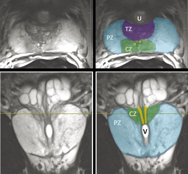
prostate zonal anatomy
Introduction to Prostate MRI Protocols: Hardware, T2-Weighted Imaging. 9 Images about Introduction to Prostate MRI Protocols: Hardware, T2-Weighted Imaging : Prostate Cancer Imaging | Radiology Key, Anatomic Imaging of the Prostate and also Normal 3T MR Anatomy of the Prostate Gland and Surrounding Structures.
Introduction To Prostate MRI Protocols: Hardware, T2-Weighted Imaging
 radiologykey.com
weighted spectroscopy protocols radiology
Prostate Anatomy
 www.aboutcancer.com
prostate bladder anatomy neck
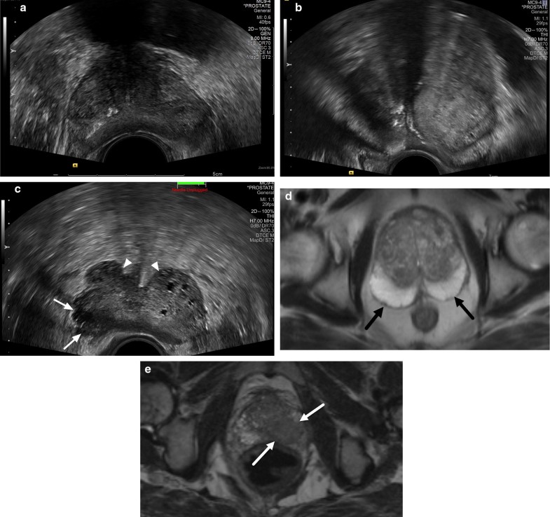
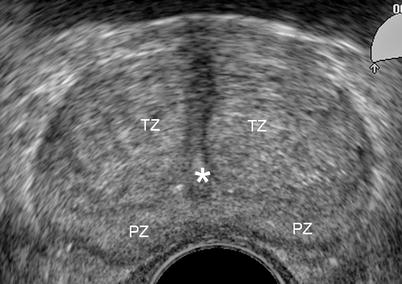
Prostate Cancer Detection And Diagnosis: Role Of Ultrasound With MRI
 link.springer.com
prostate ultrasound cancer mri anatomy diagnosis detection role fig correlates calcifications
Anatomic Imaging Of The Prostate
 www.hindawi.com
ejaculatory gland seminal zonal ducts vesicles stroma anatomic fibromuscular afs
Current Status Of Transrectal Ultrasound-guided Prostate Biopsy In The
 www.clinicalradiologyonline.net
transrectal biopsy guided diagnosis prostatic
Prostate Cancer Imaging | Radiology Key
 radiologykey.com
prostate cancer ultrasound anatomy imaging trus transrectal normal radiology sonography study key career medical college fig facts radiologykey
Scrotum Flashcards | Easy Notecards
 www.easynotecards.com
prostatic zone portion ducts urethra seminal enter where scrotum easynotecards
Normal 3T MR Anatomy Of The Prostate Gland And Surrounding Structures
 www.hindawi.com
3t surrounding
Radiology Anatomy Images : Brain Sagittal MRI Anatomy Thalamus And Cortex
 radiology-anatomy.blogspot.com
mri anatomy prostate gland penis radiology sagittal adjacent critical bladder rectum structures several including
Radiology anatomy images : brain sagittal mri anatomy thalamus and cortex. Ejaculatory gland seminal zonal ducts vesicles stroma anatomic fibromuscular afs. Prostatic zone portion ducts urethra seminal enter where scrotum easynotecards