radiographic anatomy nasal bone
PPT - Normal Radiographic Anatomy of the Equine Hind Limb PowerPoint. 9 Pictures about PPT - Normal Radiographic Anatomy of the Equine Hind Limb PowerPoint : subnasale – Dr. G's Toothpix, Dentistry lectures for MFDS/MJDF/NBDE/ORE: Anatomical Landmarks Of and also subnasale – Dr. G's Toothpix.
PPT - Normal Radiographic Anatomy Of The Equine Hind Limb PowerPoint
 www.slideserve.com
equine tarsus normal anatomy hind radiographic limb ppt powerpoint presentation
Subnasale – Dr. G's Toothpix
 drgstoothpix.com
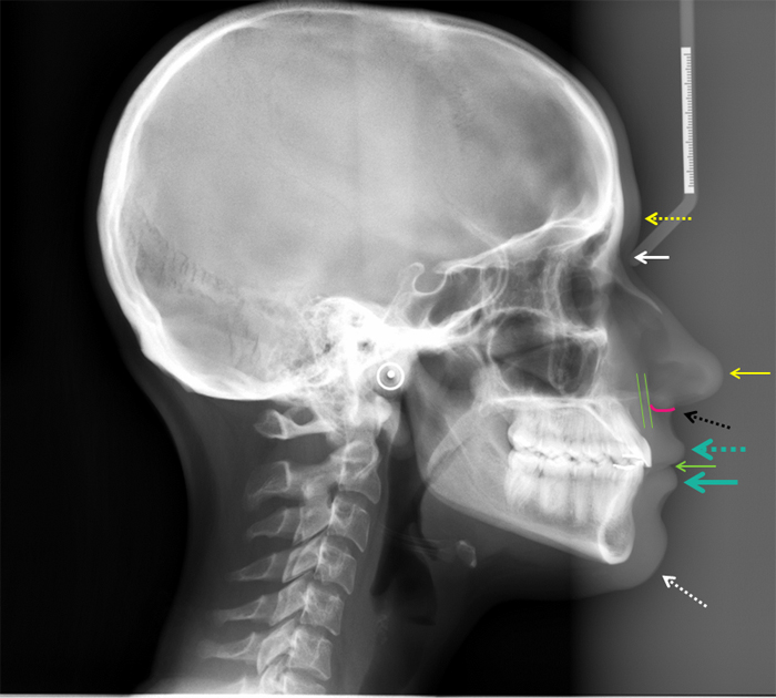
lateral skull anatomy cephalometric glabella radiograph soft tissue point process radiographic cephalometrics something looking radiographs interpretation drgstoothpix
Normal Radiographic Anatomical Landmarks
 www.slideshare.net
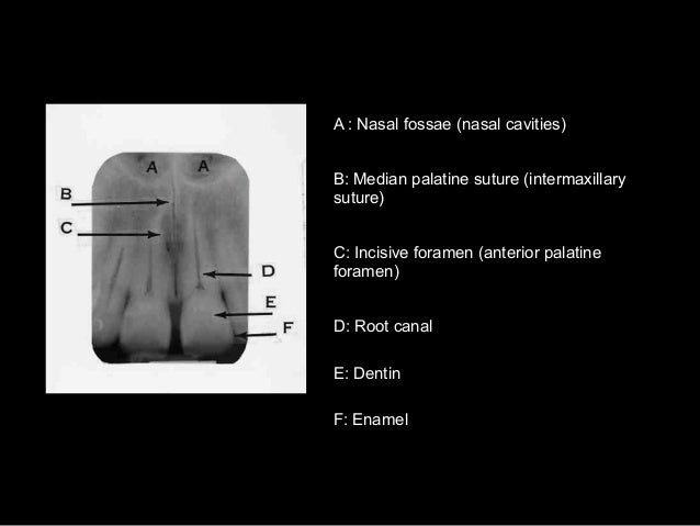
anatomical radiographic anatomic suture median palatine maxillary palatal periapical radiographs nasal radiolucent septum sinus
Normal Radiographic Anatomy
 www.slideshare.net
radiographic anatomy suture palatine median incisive intermaxillary
Inferior Nasal Concha – Dr. G's Toothpix
 drgstoothpix.com
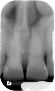
nasal concha inferior periapical radiograph incisor central radiographs radiographic
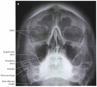
FACIAL BONES X RAY | PARIETOACANTIAL PROJECTION - RadTechOnDuty
 radtexts.blogspot.com
method
Anatomy On Radiographs: Intraoral Radiographs Part 2 – Dr. G's Toothpix
 drgstoothpix.com
mandibular nerve inferior alveolar radiographs intraoral premolar radiolucent
Dentistry Lectures For MFDS/MJDF/NBDE/ORE: Anatomical Landmarks Of
 dentallecnotes.blogspot.ca
anatomical radiograph dentistry mfds mjdf nbde lectures
Normal Radiographic Anatomical Landmarks
 www.slideshare.net
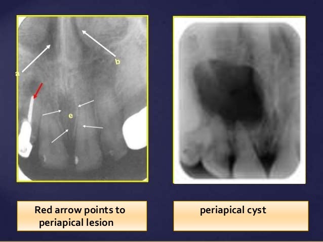
radiographic anatomical periapical cyst
Radiographic anatomical periapical cyst. Nasal concha inferior periapical radiograph incisor central radiographs radiographic. Anatomy on radiographs: intraoral radiographs part 2 – dr. g's toothpix