sinus bone anatomy
Bones: Skull, maxilla. – Anatomy & Physiology. 9 Pictures about Bones: Skull, maxilla. – Anatomy & Physiology : Anterior View of the Face in Coronal Section at the Sphenoid Sinus, 8 – Augmentation Grafting of the Maxillary Sinus for Placement of and also Untitled Document [bio.sunyorange.edu].
Bones: Skull, Maxilla. – Anatomy & Physiology
 integrativewellnessandmovement.com
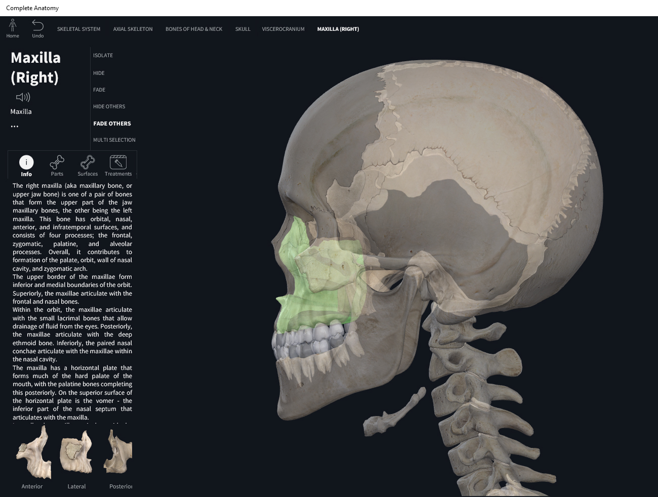
maxilla
Anterior Petrosectomy And Extended Middle Fossa Approach | Neuroanatomy
 www.neurosurgicalatlas.com
fossa
Os Sphenoidale (Keilbein) | Sphenoid Bone, Skull, Human Skull Anatomy
 www.pinterest.es
sphenoidale keilbein sphenoid sphenoidalis occipital kenhub clivus sella turcica cranium sinus
Nasal Bones Radiographic Anatomy - WikiRadiography
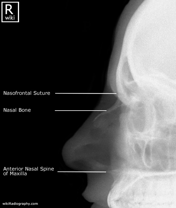 www.wikiradiography.net
nasal bones radiographic radiology radiography wikiradiography whg artrosis maxillary ehg femur anchor1
Anterior View Of The Face In Coronal Section At The Sphenoid Sinus
 www.neurosurgicalatlas.com
sinus sphenoid coronal neurosurgicalatlas
Untitled Document [bio.sunyorange.edu]
![Untitled Document [bio.sunyorange.edu]](http://bio.sunyorange.edu/updated2/comparative_anatomy/anat_3/skull_maxillary/max_cat.jpg) bio.sunyorange.edu
bio.sunyorange.edu
skull cat anatomy nasal maxillary updated2 bones evolution thinking comparative facial palate sunyorange bio edu face anat references
Anatomy On Radiographs: Intraoral Radiographs Part 2 – Dr. G's Toothpix
 drgstoothpix.com
mental foramen anatomy radiographs periapical premolar intraoral radiolucent near mandible second mesial radiographic diagram area apex round
8 – Augmentation Grafting Of The Maxillary Sinus For Placement Of
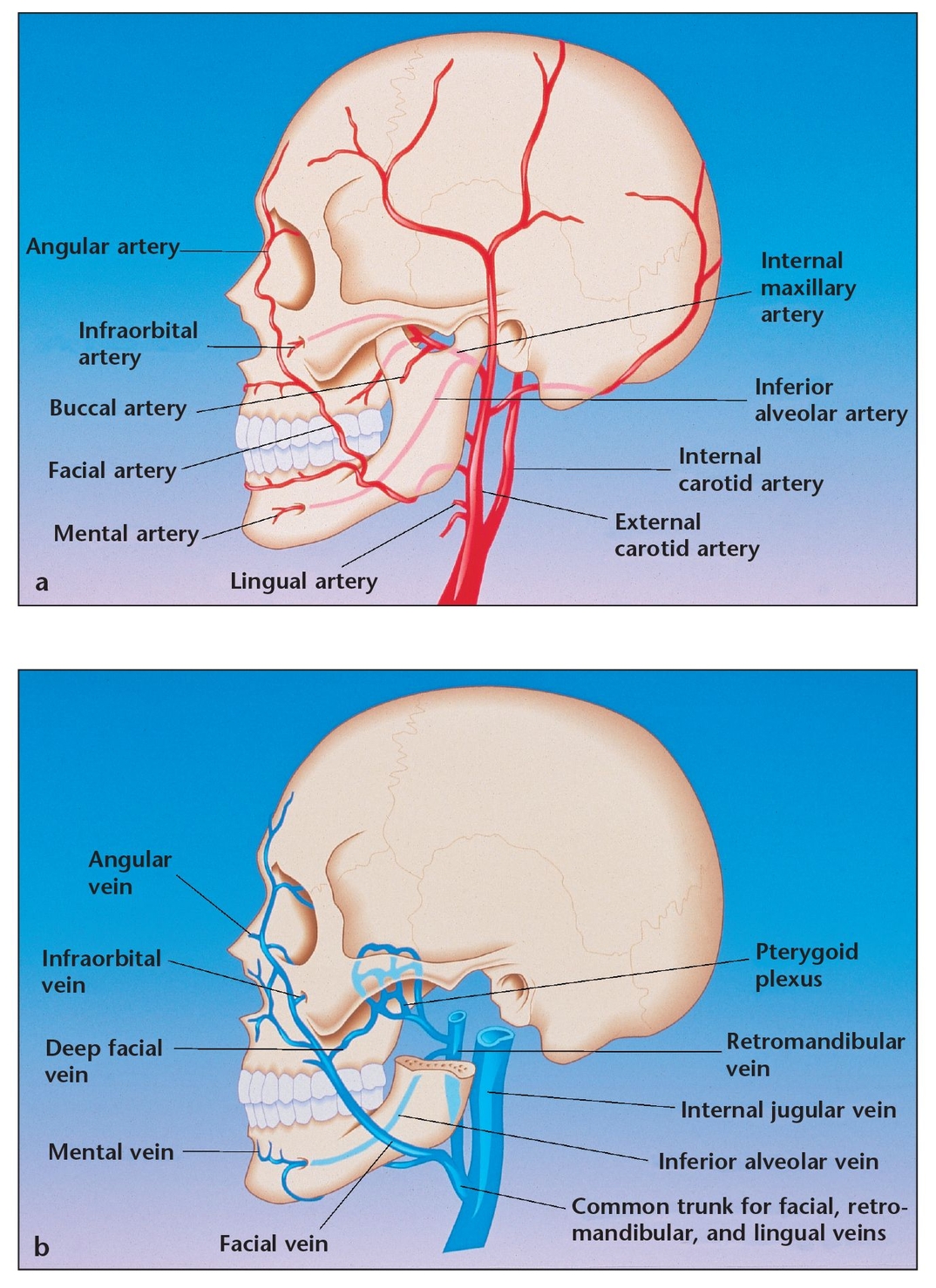 pocketdentistry.com
sinus maxillary artery facial dental anatomy drainage supply blood venous area carotid
Superior View Of The Right Orbit | Neuroanatomy | The Neurosurgical
 www.neurosurgicalatlas.com
orbit superior right neuroanatomy rhoton correlation surgical neurosurgicalatlas
8 – augmentation grafting of the maxillary sinus for placement of. Mental foramen anatomy radiographs periapical premolar intraoral radiolucent near mandible second mesial radiographic diagram area apex round. Os sphenoidale (keilbein)