sinus ct anatomy
Atrophy of the Posterior Cricoarytenoid Muscle as an Indicator of. 9 Pics about Atrophy of the Posterior Cricoarytenoid Muscle as an Indicator of : Image | Radiopaedia.org, Radiographic Anatomy of the Orbit and Visual Pathways | Radiology Key and also CT anatomy of Neck Spaces RV.
Atrophy Of The Posterior Cricoarytenoid Muscle As An Indicator Of
 www.ajnr.org
muscle cricoarytenoid laryngeal palsy ventricle nerve posterior atrophy neck head paralysis arytenoid quizlet recurrent indicator ajnr fig scc
Normal Anatomy Of The Base Of The Skull, Orbit, Pituitary And Cranial
 pn.bmj.com
cranial pituitary bmj pn
Congenital And Acquired Conditions Of The Aortic Root: Multidetector
 pmj.bmj.com
aortic
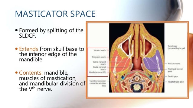
CT Anatomy Of Neck Spaces RV
 pt.slideshare.net
masticator mandible
Image | Radiopaedia.org
 radiopaedia.org
sinus sagittal superior radiopaedia vein cerebral angiogram radiology sinuses ventricles meninges confluence annotated
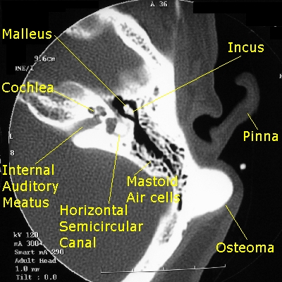
Osteoma CT - Wikidoc
 www.wikidoc.org
ct osteoma mastoid scan temporal wikidoc axial bones showing left labeled
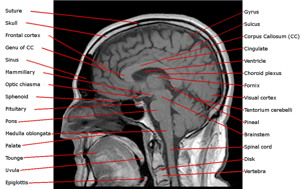
Annotated Sagittal T1 Midline MRI Scan Of Reigh's Brain | Flickr
 www.flickr.com
brain mri sagittal scan t1 midline annotated anatomy dissection reigh quiz vita parts ap psych sheep c1 english
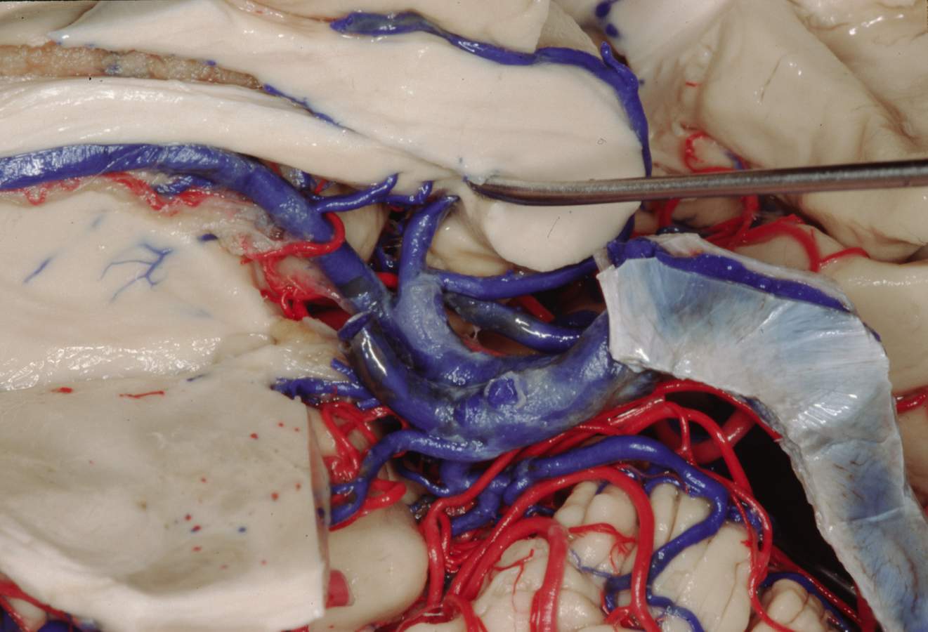
Medial View Of The Deep Cerebral Veins | Neuroanatomy | The
 www.neurosurgicalatlas.com
cerebral veins deep correlation surgical medial neurosurgicalatlas
Radiographic Anatomy Of The Orbit And Visual Pathways | Radiology Key
 radiologykey.com
anatomy orbit pathways radiographic visual bone
Anatomy orbit pathways radiographic visual bone. Ct osteoma mastoid scan temporal wikidoc axial bones showing left labeled. Medial view of the deep cerebral veins